mri protocols and planning pdf
MRI Protocols and Planning: A Comprehensive Overview
Comprehensive MRI planning involves standardized protocols, patient safety, and optimized image reconstruction techniques, as detailed in recent guidelines from organizations like the AHA/ASA.
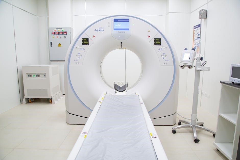
Developing robust MRI protocols is crucial for diagnostic accuracy and efficient workflow. This process necessitates a deep understanding of the clinical indication, patient factors, and the capabilities of the MRI system. Standardized protocols, often found in resources like the UNC Pediatric Body MRI Protocol Manual, ensure consistency and comparability of results.
Early management of conditions like stroke benefits from focused abbreviated surveys, as highlighted in RadioGraphics guidelines. Protocol development also involves careful consideration of contrast agent administration, utilizing agents like Diamox in neuroimaging, and optimizing image reconstruction for clarity. The CMSC is actively updating MS MRI guidelines, demonstrating the dynamic nature of protocol refinement.
Importance of Standardized Protocols
Standardized MRI protocols are paramount for ensuring consistent image quality and reliable diagnoses. They minimize variability between technologists and scanners, facilitating accurate interpretation and longitudinal follow-up. Guidelines from organizations like the AHA/ASA emphasize their role in acute stroke imaging, enabling rapid assessment with protocols like the six-minute MRI.
Protocols, such as those for pediatric MRI, detailed in the UNC manual, address specific patient needs and positioning. Updates to MS MRI guidelines from the CMSC further illustrate the ongoing effort to refine and standardize practices. Consistent protocols streamline workflow and reduce the potential for errors, ultimately improving patient care.
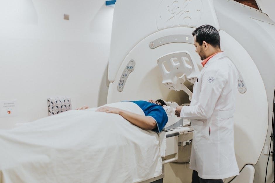
Patient Preparation and Safety Considerations
Thorough patient preparation is crucial for safe and effective MRI examinations. This begins with rigorous screening for contraindications, ensuring no metallic implants or devices pose a risk. Patient positioning and immobilization are vital, particularly in pediatric cases, as highlighted by UNC guidelines emphasizing hand placement within the field of view.
Protocols often involve pre-scan medication, like Diamox for brain perfusion studies, requiring a waiting period before imaging. Clear communication and addressing patient anxiety are essential. Adherence to safety protocols minimizes risks and optimizes image quality, contributing to accurate diagnoses.
Screening for Contraindications
Meticulous screening is paramount before any MRI scan. This involves a detailed questionnaire and physical assessment to identify absolute and relative contraindications. Absolute contraindications include metallic implants like pacemakers, cochlear implants, and certain aneurysm clips. Relative contraindications, requiring careful evaluation, encompass pregnancy, renal insufficiency (due to contrast risk), and severe claustrophobia.
Thorough documentation of all implants and medical history is essential. Protocols must address potential risks and alternative imaging modalities if contraindications exist, ensuring patient safety remains the top priority.
Patient Positioning and Immobilization
Optimal patient positioning is crucial for image quality and diagnostic accuracy. Supine positioning is common, but specific protocols dictate adjustments for different anatomical regions. Immobilization minimizes motion artifacts, particularly vital for neuro and spine imaging. Pediatric patients require specialized positioning, such as hands splayed under legs to ensure full FOV coverage.
Proper head support and body coils are essential. Protocols should detail coil selection and positioning techniques. Clear communication with the patient regarding the need for stillness is paramount, alongside offering comfort and reassurance throughout the scan.
Neuro MRI Protocols
Neuro MRI protocols encompass a range of sequences for comprehensive brain and spine evaluation. Routine brain protocols often include sagittal T1, axial DWI, axial T1, axial T2 FLAIR, and axial T2 weighted imaging. Perfusion imaging, utilizing contrast agents, is frequently incorporated, sometimes with a half-dose technique following Diamox administration to assess cerebral blood flow.
Specific protocols are tailored for acute stroke assessment, emphasizing rapid acquisition times. These protocols aim to differentiate ischemic core from penumbral tissue, guiding treatment decisions. Updated guidelines continually refine these sequences for improved diagnostic yield.
Routine Brain MRI Protocol
A standard brain MRI protocol typically begins with sagittal T1-weighted imaging for anatomical orientation. This is followed by axial T2-weighted and FLAIR sequences to detect subtle pathology. Diffusion-weighted imaging (DWI) is crucial for identifying acute ischemic changes, while axial T1-weighted images assess for hemorrhage or structural abnormalities.
Perfusion imaging, often performed with contrast, evaluates cerebral blood flow dynamics. The protocol may be adjusted based on clinical indication, with additional sequences like gradient echo or susceptibility-weighted imaging added as needed for specific assessments.
Stroke Imaging Protocols
Rapid MRI is paramount in acute stroke evaluation. Protocols prioritize swift detection of ischemic changes using DWI and FLAIR sequences. Six-minute MRI protocols are emerging, pushing the boundaries of speed without compromising diagnostic quality. Perfusion imaging, utilizing contrast agents, assesses penumbral regions and guides treatment decisions.
Guidelines from the AHA/ASA emphasize the importance of MRI in acute ischemic stroke management. Protocols often include pre- and post-contrast imaging to differentiate between ischemic and hemorrhagic events, crucial for determining appropriate interventions.
Acute Ischemic Stroke MRI
The core of acute ischemic stroke MRI focuses on Diffusion-Weighted Imaging (DWI), revealing areas of restricted diffusion within minutes of symptom onset. FLAIR sequences complement DWI, highlighting edema and potentially identifying larger infarcts. Rapid acquisition is critical, often employing echo-planar imaging (EPI) techniques.
Protocols frequently include T1-weighted imaging to exclude hemorrhage and assess for early signs of blood-brain barrier disruption. Contrast-enhanced MRI can delineate the penumbra, but timing is crucial to avoid obscuring early ischemic changes. The six-minute protocol aims for expedited diagnosis.
Perfusion Imaging in Stroke
Perfusion MRI assesses cerebral blood flow (CBF), CBV, and MTT, identifying the ischemic penumbra – tissue at risk of infarction. Dynamic Susceptibility Contrast (DSC) perfusion is commonly used, employing a rapid injection of contrast agent followed by serial imaging. Post-processing generates perfusion maps, visualizing areas of reduced blood flow.
Perfusion data aids in patient selection for thrombolysis or thrombectomy, guiding treatment decisions based on the extent of salvageable tissue. Protocols often involve a half-dose contrast injection, as noted in neuro MRI updates. Combining DWI and perfusion provides a comprehensive assessment of stroke severity and potential for recovery.
Spine MRI Protocols
Spine MRI protocols require careful consideration of the region of interest – cervical, thoracic, or lumbar – and the clinical indication. Sagittal T1 and T2 weighted images are fundamental for assessing spinal alignment and morphology. Axial imaging complements sagittal views, visualizing disc herniations and foraminal stenosis.
STIR sequences are valuable for detecting edema associated with inflammation or trauma. Contrast-enhanced imaging can delineate nerve root compression or spinal cord lesions. Protocol optimization minimizes scan time while maintaining diagnostic image quality, crucial for patient comfort and workflow efficiency.
Routine Spine Protocol
A routine spine protocol typically begins with sagittal T1 and T2 weighted images, providing anatomical detail. Axial T2 weighted images are then acquired to assess disc pathology and nerve root compression. Sagittal STIR sequences are essential for detecting edema, indicating inflammation or recent injury.
For contrast-enhanced studies, T1 weighted images are obtained pre- and post-gadolinium administration. This aids in identifying areas of abnormal enhancement, such as tumors or infections. Image parameters, including slice thickness and field of view, should be standardized for consistent results and accurate comparisons.
Pediatric MRI Protocols
Pediatric MRI requires specialized protocols due to unique physiological and developmental considerations. Positioning is crucial; UNC Pediatric Body MRI protocols emphasize splayed arms under the legs to ensure complete FOV coverage. Minimizing scan time is paramount to reduce the need for sedation, especially in younger patients.
Age-appropriate communication and preparation are essential to alleviate anxiety. Lowering field strength when feasible can reduce specific absorption rate (SAR) concerns. Pediatric radiologists should review all cases of patients 18 years and under, ensuring accurate interpretation and tailored reporting.
Specific Considerations for Pediatric Patients
Pediatric patients necessitate careful attention to radiation safety and minimizing scan times. Sedation protocols must be individualized, considering age, weight, and medical history. Communication is key; explain procedures in child-friendly terms to reduce anxiety and ensure cooperation. Motion artifact reduction is vital, employing techniques like fast sequences and breath-holding instructions when appropriate.
Contrast agent administration requires precise dosage calculations based on weight. Monitoring for adverse reactions is crucial. Parental presence during the scan can provide comfort and support. Adapting protocols to the child’s developmental stage optimizes image quality and patient experience.
Positioning Guidelines for Pediatric Body MRI
Proper positioning is paramount for comprehensive pediatric body MRI. Arms should be positioned splayed open under the legs, maintaining straight alignment to ensure full upper extremity coverage within the field of view (FOV). Stitched images are valuable for confirming adequate coverage and hand positioning.
Always verify the case with a pediatric reading MD, as patients 18 years and under typically require a total body protocol, potentially including a separate brain MRI. Immobilization is crucial to minimize motion artifacts, utilizing appropriate padding and support. Careful attention to anatomical landmarks ensures accurate image interpretation.
Multiple Sclerosis (MS) MRI Protocols
Ongoing updates refine magnetic resonance imaging (MRI) guidelines for multiple sclerosis (MS) patients. The Consortium of Multiple Sclerosis Centers (CMSC) protocol is frequently revised to incorporate advancements in imaging techniques and diagnostic criteria. These updates aim to enhance the detection of disease activity and monitor treatment response effectively.

Standardized protocols are essential for consistent and comparable results across different centers. Current recommendations emphasize the importance of specific sequences, such as T1-weighted, T2-weighted, and FLAIR imaging, to visualize lesions and assess disease progression accurately.
Updates to MS MRI Guidelines
Proposed revisions to MS MRI guidelines focus on optimizing lesion detection and characterization. These updates address evolving understandings of disease pathology and the need for more sensitive imaging biomarkers. Emphasis is placed on incorporating advanced techniques, such as magnetization transfer imaging and diffusion tensor imaging, to assess subtle disease changes.
The goal is to improve diagnostic accuracy and personalize treatment strategies. Updated protocols also consider the role of MRI in monitoring disease-modifying therapies and predicting long-term outcomes. Collaboration between radiologists and neurologists is crucial for implementing these changes effectively.
Rectum MRI Protocols
Rectal MRI protocols typically employ a 1.5T scanner with a phased-array surface coil, positioned to ensure complete coverage of the anal canal and distal rectum. Sagittal and axial T2-weighted images are essential for anatomical assessment, while diffusion-weighted imaging (DWI) aids in differentiating benign from malignant lesions. Post-contrast T1-weighted sequences further enhance lesion characterization.
Careful patient positioning – supine with appropriate coil placement – is critical for image quality. Protocols may include dynamic contrast-enhanced imaging to evaluate tumor vascularity. Standardized reporting templates ensure consistent interpretation and facilitate communication between radiologists and clinicians.
Protocol Details for Rectal Imaging
A typical rectal MRI protocol on a 1.5T Philips system begins with axial and sagittal T2-weighted sequences, providing excellent soft tissue contrast. Subsequent axial DWI with corresponding ADC maps are crucial for lesion detection. T1-weighted images, both pre- and post-gadolinium contrast, are then acquired to assess for enhancement patterns.
Specific parameters include slice thickness of 4-5mm, with a field of view tailored to the patient’s anatomy. Contrast administration usually involves gadolinium-based agents, with dosage adjusted based on patient weight and renal function. High-resolution imaging is vital for accurate staging and treatment planning.
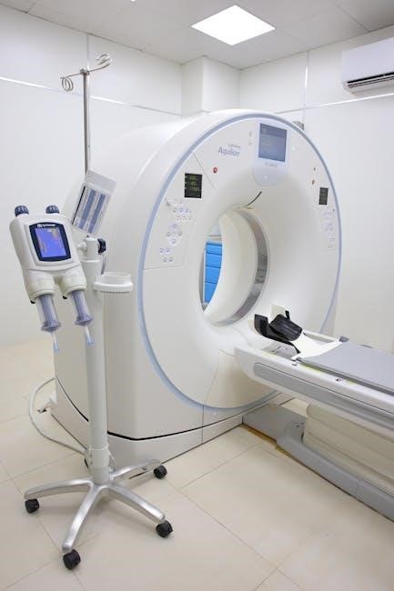
Contrast Agent Administration
Contrast agents, primarily gadolinium-based, significantly enhance MRI visualization, improving lesion detection and characterization. Types include macrocyclic and linear agents, with macrocyclic generally preferred due to reduced risk of nephrogenic systemic fibrosis (NSF). Dosage is typically calculated based on patient weight, ranging from 0.1 to 0.2 mL/kg.
Injection parameters are critical; a power injector ensures consistent delivery at a rate of 2-5 mL/second, followed by a saline flush. Careful monitoring for adverse reactions, such as allergic responses, is essential. Renal function assessment is paramount before administration.
Types of Contrast Agents Used
Gadolinium-based contrast agents (GBCAs) are the mainstay for MRI enhancement, categorized into linear and macrocyclic compounds. Macrocyclic agents, like gadoteridol and gadobutrol, exhibit greater stability and lower risk of NSF compared to linear agents such as gadodiamide;
Recent considerations focus on minimizing GBCA retention, particularly with repeated administrations. Gadoxetate disodium (Primovist), a hepatobiliary agent, is utilized for liver imaging. Iron oxide nanoparticles serve as negative contrast agents, useful in liver and spleen assessments.
Dosage and Injection Parameters
Contrast agent dosage varies based on the clinical indication, patient weight, and agent type. Typically, GBCAs are administered at 0.1-0;2 mmol/kg body weight. Injection rates are crucial; slower rates (e.g., 5 mL/second) are preferred for perfusion studies to accurately assess cerebral blood volume.
For brain perfusion, protocols often utilize a half-dose technique, with Diamox administration preceding contrast injection. Saline flush before and after contrast injection ensures complete delivery. Monitoring for adverse reactions is paramount, with immediate access to emergency equipment.
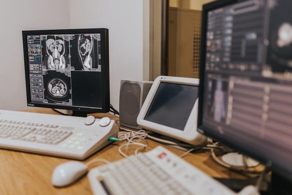
Image Reconstruction and Post-Processing
Optimizing image quality requires careful selection of reconstruction kernels and filtering techniques. Techniques like minimum intensity projection (mIP) and maximum intensity projection (mIP) enhance visualization of vascular structures. Advanced post-processing, including multiplanar reconstruction (MPR) and 3D rendering, aids in detailed anatomical assessment.
Stitching images, particularly in pediatric body MRI, improves field-of-view coverage. Perfusion imaging benefits from specialized software for calculating cerebral blood volume, flow, and mean transit time. Precise image analysis is vital for accurate diagnosis and treatment planning.
Optimizing Image Quality
Achieving superior image quality necessitates meticulous attention to several parameters. Selecting appropriate reconstruction kernels—such as bone, soft tissue, or high-resolution—significantly impacts image sharpness and noise levels. Careful filtering minimizes artifacts while preserving crucial anatomical details.
FOV and matrix size adjustments are critical for balancing spatial resolution and scan time. Proper shimming ensures magnetic field homogeneity, reducing geometric distortions. Optimized slice thickness impacts signal-to-noise ratio and image clarity. Regular quality control is essential for maintaining scanner performance.
Advanced Post-Processing Techniques
Beyond basic reconstruction, advanced post-processing enhances diagnostic capabilities. Perfusion mapping, utilizing techniques like dynamic susceptibility contrast (DSC), visualizes cerebral blood flow, crucial in stroke assessment. Diffusion tensor imaging (DTI) reveals white matter tract integrity, aiding in neurological evaluations.
Multiplanar reconstruction (MPR) and maximum intensity projection (MIP) facilitate visualization in complex anatomical regions. 3D rendering provides immersive anatomical views. Quantitative analysis, including volumetry and lesion burden assessment, offers objective measurements. These techniques demand specialized software and expertise for accurate interpretation.